Date/Time
15th Dec 2021
3pm BST | 4pm CEST | 7am PDT | 10am EDT
Event type
Webinar
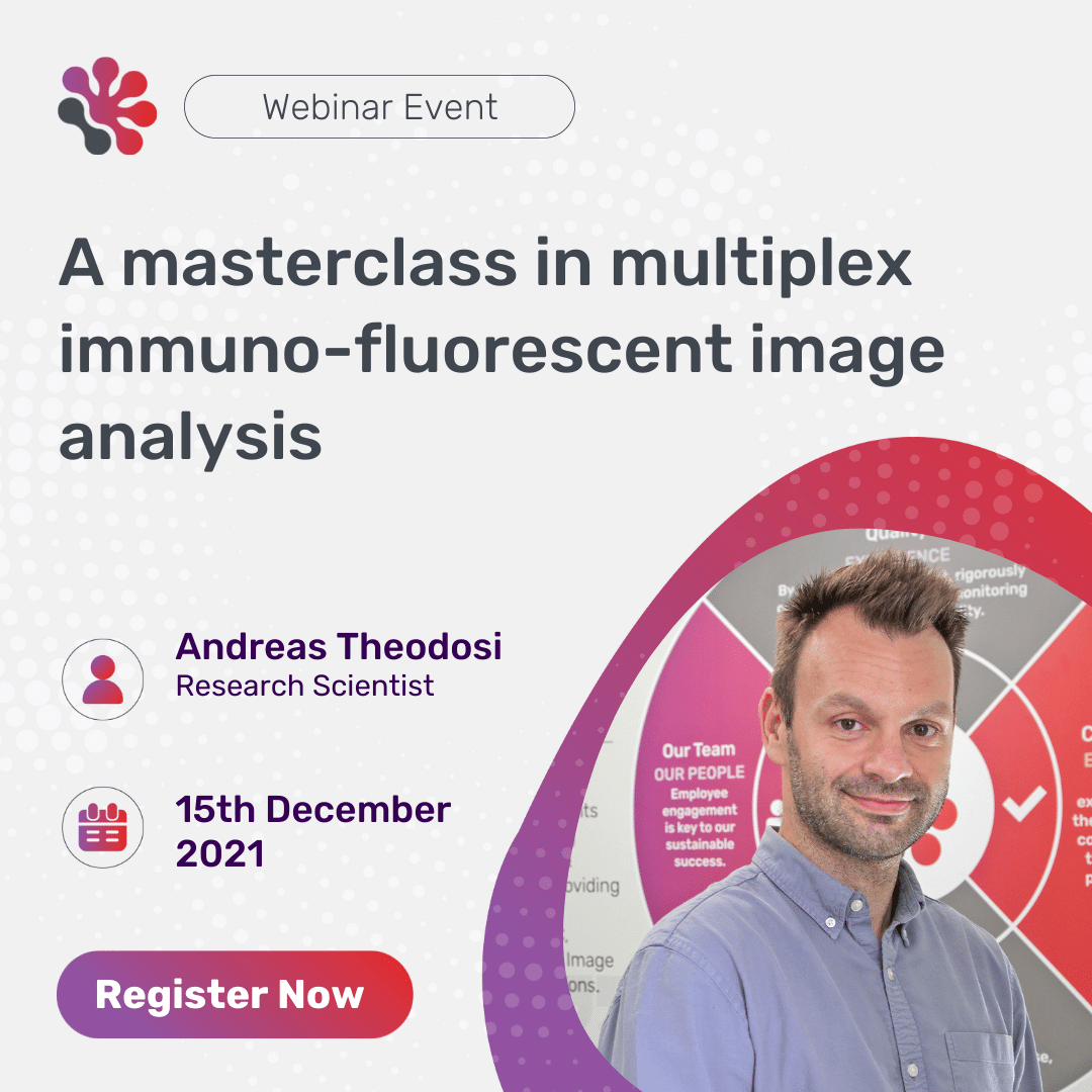
Speaker
Andreas Theodosi
Immuno-fluorescent multiplexing is a powerful tool in the field of tumor immunology for precise quantification of protein expression, distribution, and activation in situ.
In this expert webinar, join our research scientist Andreas Theodosi as he looks at the image analysis of multiplexed immune-fluorescent stained tumor samples. Theodosi will discuss the benefits and limitations of using fluorescent multiplexing over other methods such as chromogenic single, serial section or chromogenic multiplexed techniques.
Key learning objectives:
- The differences between different image analysis techniques
- The workflow of immuno-fluorescent multiplexing
- The analysis capabilities and quantitative endpoints available when working with immuno-fluorescent multiplexed images
Speakers

Andreas Theodosi
Research Scientist
Andreas graduated from University of Hull with BSc (Hons) 1st class in biomedical science in 2015. After graduating, Andreas became a course leader and a member of the Learning Technology and Innovation Forum at Burton and South Derbyshire College. In 2018, Andreas joined HistologiX Ltd as a research scientist working across histology, immunohistochemistry and digital image analysis. He has a particular interest in computer-based systems, including Indica Lab’s HALO 3.0, Hamamatsu Nanozoomer with NDP Viewer, Zeiss Axio Scan with Zen, Leica Bond, Roche Benchmark & Discovery Ultra, Microsoft Excel with Visual Basics, and Haeir Monitoring System with real-time reporting software.