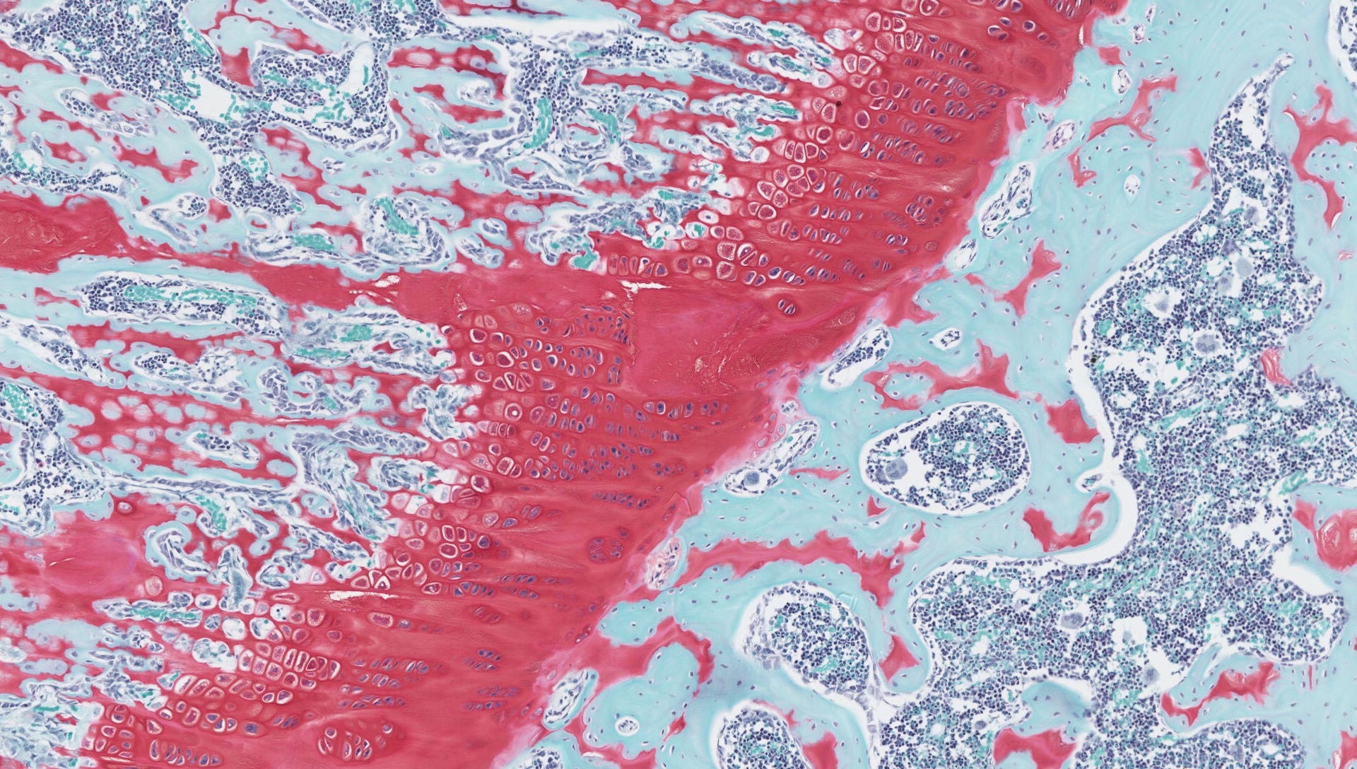
Using our Hamamatsu NanoZoomer v2 and Zeiss Axioscan Z1 HistologiX can provide brightfield and fluorescent digital scanning services. Scanning at magnifications of x20 and x40, we generate high quality images of your glass slides to be used for archiving or digital image analysis. We will upload these onto a secure Egnyte™ cloud server where you can remotely access these images in an instant from your office.
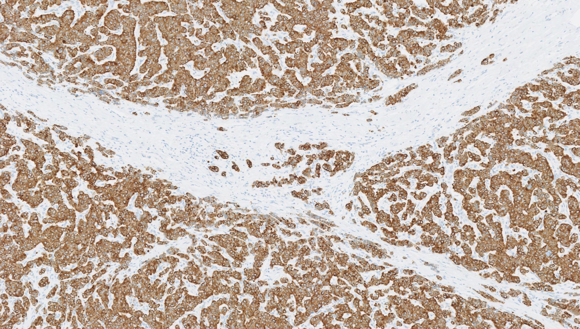
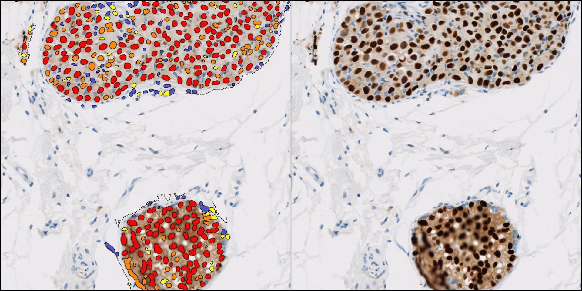
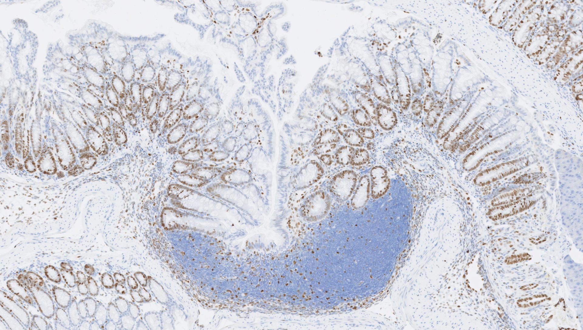
With our gold standard image analysis platform, HALO™ from Indica Labs, HistologiX is equipped to support your quantitative tissue analysis requirements. HALO™ is an intuitive image analysis platform for digital pathology which reports morphological and multiplexed expression data on a cell-by-cell basis across entire tissue sections.
Combining histology analytics, interactive results and a plethora of analysis algorithms, HALO™ allows cell based, area based and spatial quantification of biomarkers within target tissues. Using the tissue classifier module tissue segmentation can be automated. Together this powerful tool set supports analysis across a wide range of applications such as oncology, neuroscience, metabolic disorders, toxicological pathology, amongst others. Designed to handle huge data sets from IHC, IF and ISH experiments, results can be presented as a detailed dataset, percentage of total cells present, or as an H-Score. With over 20 years experience in this field, our experts are uniquely placed to deliver analysis solutions for your studies along with interpretive comments.
Stay up to date with HistologiX