Unfolding the Mystery of IHC vs IF
The tale of modern medicine would be incomplete without the mention of the revolutionary tools that researchers employ in their quest to unravel the complex mechanisms of life and disease. Among these, two techniques stand out – Immunofluorescence (IF) and Immunohistochemistry (IHC).
These methodologies, each with its unique prowess, help scientists visualise the intricate dance of proteins within cells and tissues. Now, if you’re wondering, “What’s the big deal about Immunofluorescence vs IHC?”, hold onto that curiosity as we navigate through the captivating world of these lab techniques. To discover more about how these fascinating techniques are applied in a professional lab, visit our Cellular Pathology services.
Immunofluorescence – A Vivid Palette of Molecular Recognition
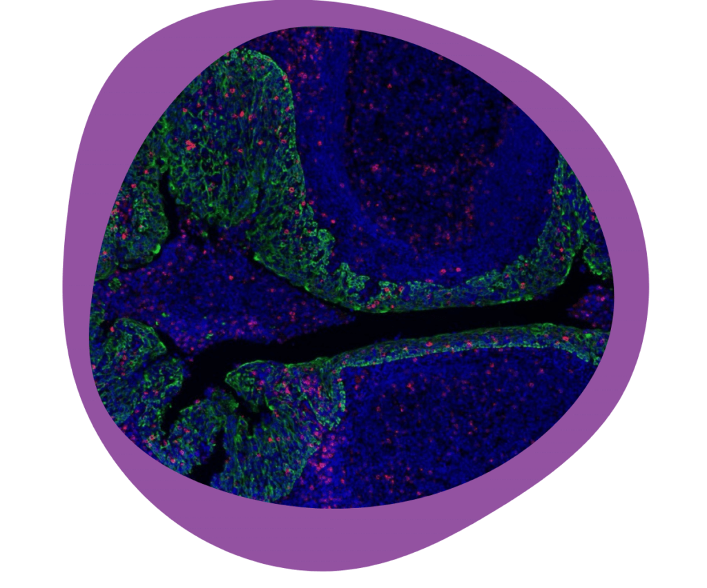
Immunofluorescence, or IF, is a technique that harnesses the specificity of antibodies to target proteins and then uses the dazzling world of fluorescence to visualise them.
The Art and Science Behind Immunofluorescence
How does Immunofluorescence work? In the heart of it all is the relationship between antibodies and antigens. With antibodies acting as unique ‘guides,’ leading us straight to our target protein.
Immunofluorescence: More Than Meets the Eye
Direct and indirect IF are the two primary types. The former uses a single, fluorescently labelled antibody, while the latter utilises a secondary antibody to amplify the signal. So, why might you use one or the other? Well, direct IF is quicker and reduces non-specific binding. In contrast, indirect IF offers higher sensitivity – a classic case of speed vs sensitivity!
The Scope of Immunofluorescence in Biomedical Research
From investigating cell signalling pathways to diagnosing infectious diseases, IF has revolutionised many areas. Would you believe that it even plays a significant role in the COVID-19 pandemic, helping researchers understand the virus better? If you’re curious about how immunofluorescence is used in cutting-edge cancer research, learn more about our Immuno-Oncology services.
Immunohistochemistry – Painting a Detailed Picture of Cellular Life
Now that we’ve explored the world of IF, it’s time to acquaint ourselves with another heavyweight in this arena – Immunohistochemistry or IHC.
Immunohistochemistry: The Nuts and Bolts
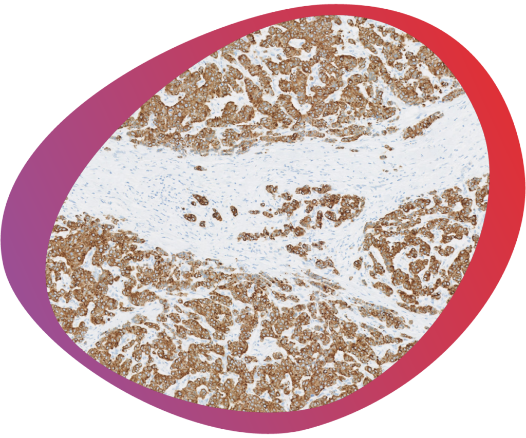
So, how does IHC work? Well, like IF, it exploits the antigen-antibody interaction. The difference, however, lies in the visualisation step. Instead of fluorescent labels, IHC uses enzymes that produce coloured reactions when exposed to specific substrates.
Unlocking the Doors to Multiple IHC Variants
Like IF, IHC isn’t a one-size-fits-all technique. There are multiple variants, including direct, indirect, and multiple staining IHC. Each comes with its pros and cons, addressing unique research questions.
IHC: A Powerful Tool in Diagnostics and Beyond
What are the applications of IHC? From diagnosing cancers to investigating neurological disorders, IHC has left indelible marks in the world of biomedical science. Without it, we would lack the insights we currently hold about various diseases.
IHC vs IF – Weighing the Differences and Similarities
With a basic understanding of both IF and IHC under our belts, let’s delve into the heart of our topic – Immunofluorescence vs IHC.
The Similarities: More Than What Meets the Eye
IF and IHC might seem distinct, but they share more than you’d initially think. Both techniques rely on the power of antibodies to target specific proteins, both utilise signal amplification methods, and both offer the ability to study cells and tissues with high specificity.
The Distinctions: It’s All in the Details
Now, let’s look at how IF and IHC are different? The primary distinction lies in the visualisation method – fluorescence for IF and chromogenic reactions for IHC. This difference paves the way for unique advantages and disadvantages, which we’ll explore next.
Advantages and Limitations of Immunofluorescence
The Strengths of Immunofluorescence
IF offers several advantages, including multi-colour imaging and live cell imaging capabilities. But what steals the show is its high resolution, capturing breath taking images of cellular life.
The Limitations of Immunofluorescence
While IF offers significant benefits, it isn’t without its limitations. The need for specialised expensive equipment for fluorescence microscopy could be a stumbling block for some labs. We have this equipment. Additionally, the fluorophores can fade over time, a phenomenon known as photobleaching. This means often it needs to be reviewed rapidly before degradation occurs and stored appropriately.
Advantages and Limitations of Immunohistochemistry
The Strengths of Immunohistochemistry
IHC scores with its long-lasting staining, compatibility with routine histological samples, and its ability to provide a permanent record of staining. But perhaps its most significant advantage is its wide adoption in diagnostic pathology, making it a cornerstone in disease diagnosis.
The Limitations of Immunohistochemistry
As with any technique, IHC isn’t flawless. Its main limitation lies in its inability to perform multi-colour staining efficiently and the lack of high-resolution imaging when compared to IF.
Choosing the Right Technique: Immunofluorescence vs IHC
With an understanding of the pros and cons of both IF and IHC, how does one choose between them?
Making an Informed Choice
Deciding between IF and IHC depends on several factors, such as the research question at hand, the available resources, and the type of samples used. Let’s say you want high-resolution images – IF might be the better choice. But if you require a permanent record, IHC might be the better option.
The Flexibility of Combining Techniques
The beauty of science lies in its flexibility. IF and IHC aren’t mutually exclusive – in fact, many of our clients often ask us to employ both techniques to gather complementary information.
The Journey of Deciphering Immunofluorescence vs IHC
In the world of biomedical research, both Immunofluorescence and Immunohistochemistry stand as powerful tools, each with its unique strengths and challenges. While they share common ground in their fundamental principles, their differences carve their unique niches in research and diagnostics.
Thus, in the debate of Immunofluorescence vs IHC, there’s no definitive winner. Instead, the choice of technique rests on the nature of your research question and the resources at hand. By understanding these techniques better, we hope you are better equipped to navigate the fascinating world of protein visualisation.
Remember, in the end, it’s not just about picking a technique. It’s about choosing a method that best answers your research questions, leading you one step closer to your scientific breakthrough. And on this journey, both Immunofluorescence and Immunohistochemistry stand as reliable companions.
Ready to dive deeper into the world of IF and IHC? Our dedicated team at HistologiX is prepared to support your research and diagnostic needs. Explore our Cellular Pathology and Immuno-Oncology services or Contact Us today.
The main difference lies in how the techniques visualise proteins. IF uses fluorescent dyes, while IHC utilises enzymes that produce colour reactions.
Absolutely! Both techniques can often provide complementary information when used on the same sample.
IF generally offers higher resolution imaging due to the properties of fluorescent microscopy.
Yes, IHC is more widely adopted in diagnostic pathology due to its compatibility with routine histological samples and long-lasting staining.
The main limitations of IF include the phenomenon of photobleaching and the requirement for specialised fluorescence microscopy equipment.
IHC’s limitations mostly revolve around its difficulty with multi-colour staining and lower resolution imaging compared to IF.
Meet the Team

Agata Stryjewska PhD
Agata is our immunofluorescence specialist with expertise in both fluorescence and chromogenic antibody validation methods. She is currently developing bespoke multiplex immunofluorescence assays for our clients’ projects. Agata also manages our Ultivue projects, offering multiplex immunofluorescence (mIF) solutions and RNAScope projects for the detection of RNA in tissues.
Agata has a Masters degree in Biotechnology from the University of Science and Technology, Wroclaw, and a PhD from the University of Leuven. She has extensive collaborative postdoctoral research experience in neuroscience, stem cells, developmental biology and immunology.